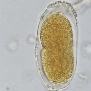
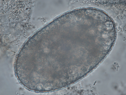
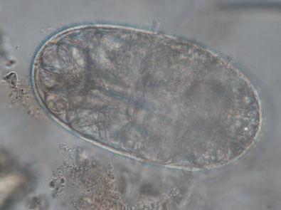
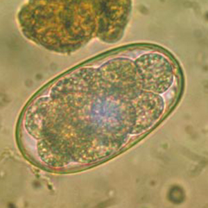
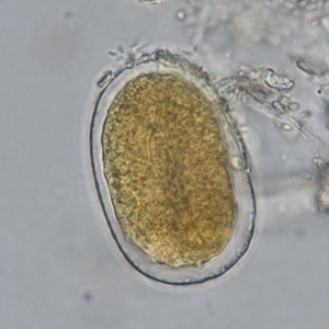
Strongyles are a group of nematodes belonging to the order Strongylida. It is a very diverse group of parasites which can affect a large number of animals. In non-human primates, strongyles belong to the Ancylostomatidae and Strongylidae families, and to the Trichostrongyloidea superfamily.
Epidemiology
Geographical distribution and species affected by strongyles depend on the genera and/or species of the parasite.
Description
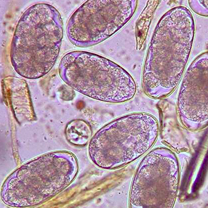
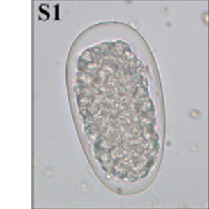
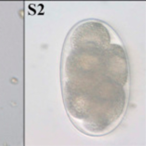
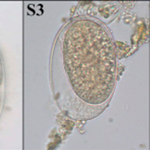
Strongyle eggs all have a similar morphology. They are oval with non-parallel lateral sides. They have a thick outer membrane and contain a morula. Their size is variable and ranges from 40 x 60 µm to 110 x 230 µm. Some of the groups above mentioned have specific characteristics which may allow their differentiation. When they remain long periods of time in the environment, strongyle eggs can contain a vermiform embryo (Garcia, 2021).
Differential diagnosis
Differential diagnosis for strongyle eggs includes parasitic and non-parasitic structures:
- Free-living nematodes: free-living nematode eggs are typically larger than strongyles, ranging from 70 to 120 µm in length and 24 to 43 µm large. Moreover, some free-living parasites have typical characteristics like refringent globules in Heterodera radicicola. Nevertheless, a coproculture or Baermann migration-based diagnosis are recommended in case of doubts.
- Strongyloides: they are included in the differential diagnosis of embryonated strongyle eggs. However, they measure 40 to 70 µm long and 20 to 35 µm large, have a very thin outer membrane and parallel lateral sides (Garcia, 2021).
- Mite eggs: they are larger than strongyle eggs, measuring 100 to 140 µm long and 50 to 80 µm large. Moreover, their content is granulomatous and contain numerous nutritive vacuoles. When the embryo is developed, the mite egg can clearly be differentiated from a strongyle egg (Petithory et al., 1995).
- Tracheid: some plant cells are of similar dimensions as strongyle eggs and can be filled with granulomatous material. However, their outer membrane is thinner, and they have a more angular shape than parasite eggs (Petithory et al., 1995).
Clinical significance
Strongyle infections typically cause gastrointestinal signs in primates. However, the significance of the infection depends largely on the parasite affecting the animal and the parasitic load (Strait et al., 2012; Calle & Joslin, 2015; Murphy, 2015).
Prophylaxis and treatment
Treatment modalities will depend on the parasite affecting the animal. However, various treatments have been described (Strait et al., 2012; Calle & Joslin, 2015):
- Thiabendazole: 50-100 mg/kg PO q24h for 1 to 2 days;
- Moxidectin: 0.5 mg/kg PO or IM;
- Mebendazole: 1550 mg/kg PO q24h for 3 to 14 days mg/kg;
- Levamisole: 10 mg/kg PO q24h for 2 to 3 days;
- Ivermectin: 0.2 mg/kg PO, IM or SC twice with 10 to 14 days of interval;
- Fenbendazole: 10-50 mg/kg PO q24h for 3 to 10 days.
