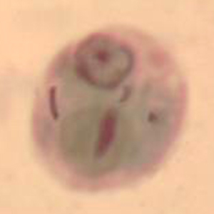
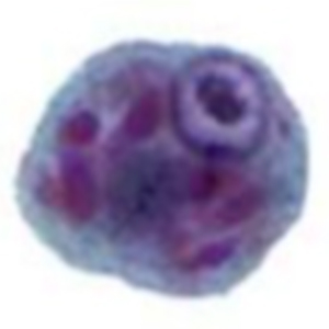
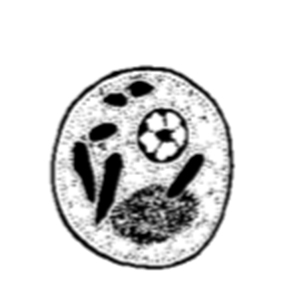
Entamoeba polecki is an amoeba affecting the cecum and colon of primates (Cogswell, 2007).
Epidemiology
Entamoeba polecki is a cosmopolitan parasite only described in Apes. It can also affect humans (Cogswell, 2007).
Description
Entamoeba polecki cysts are medium-sized (9 to 18 µm), round, and stain with Lugol. They have only one subcentral nucleus with a voluminous eccentric endosome and peripheric and granulomatous chromatin. They also contain crystalloid inclusions, which can be more easily visualized with trichrome or iron hematoxylin staining (Euzéby, 2008).
Differential diagnosis
Differential diagnosis includes medium-sized to large amoebas and non-parasitic structures. Differentiation is made based on the number of nuclei, size and visualization of different organelles with specific staining procedures. Indeed, Entamoeba coli cysts contain one to 8 nuclei, sharp-tipped crystalloid bodies, and measure 10 to 35 µm. Entamoeba histolytica cysts contain one to four nuclei, round tipped crystalloid bodies, a lipidic vacuole in their immature form, and measure 10 to 20 µm in diameter (Euzéby, 2008). Non-parasitic elements like spores do not contain intracytoplasmic structures and normally have a thicker and more refringent outer membrane (Petithory et al., 1995; Strait et al., 2012).
To summarize, an amoeba-like cyst superior to 20 µm in diameter and/or that has more than 4 nuclei will always be an Entamoeba coli. Differentiation between immature E. coli cysts, E. polecki cysts and immature E. histolytica cysts is more complicated and may require the use of specific stains like trichrome or iron hematoxylin (Euzéby, 2008).
Clinical significance
Entamoeba polecki is not pathogenic for its hosts (Cogswell, 2007; Regan et al., 2014).
Prophylaxis and treatment
As a non-pathogenic parasite, contamination by Entamoeba polecki does not require treatment. Nevertheless, hygienic measures need to be taken in case of diagnosis in a captive setting as it is considered zoonotic (Cogswell, 2007).


